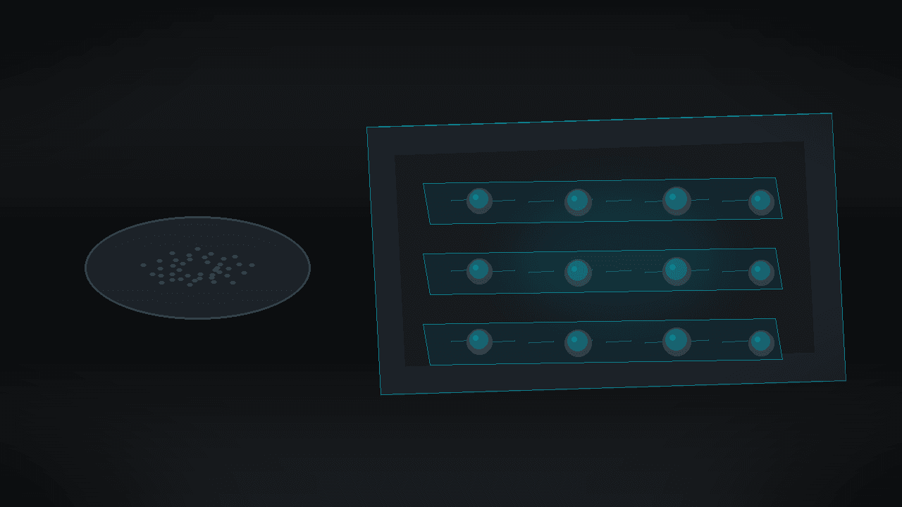
A mini-tumor in a chip reveals a drug the plate does not detect
A flat culture dish lies about a tumor more quietly than about a bacterium. Cancer cells in a classic drug test sit in a single layer, like paint on a table. A tumor in the body is a sphere: less oxygen in the core, more drug contact at the outside, and matrix plus gradients between the cells. Fluid motion adds another layer. Tissue is not a still puddle. There is a slow but steady interstitial flow. The anatomy of the problem starts here: if the lab measures a drug in flat stillness, while the patient carries a three-dimensional tumor under flow, the result can mislead long before the first clinical trial.
A team at Brazil’s Center for Research in Energy and Materials (CNPEM) in Campinas built an answer layer by layer. In ACS Measurement Science Au (DOI: 10.1021/acsmeasuresciau.6c00140), Iris Renata Sousa Ribeiro of the Brazilian Nanotechnology Laboratory (LNNano-CNPEM) and colleagues describe a reversible microfluidic chip. It grows spheroids — small, spherical cell aggregates — lets researchers retrieve them for further analysis, and tests an anticancer drug under continuous flow rather than in a standing medium.
Why flat culture fails earlier than the patient
In oncology drug development, hope that a 2D plate will say everything is fading. Spheroids and organoids better capture tumor geometry: oxygen and nutrient gradients, cell–cell contacts, and diffusional resistance into the mass. Literature has long warned that many candidates look promising in 2D and then fail in models closer to the body or in the clinic. That is not magic of a “third dimension.” It is physics and biology at the scale of a millimeter-scale sphere.
Microfluidics could add another layer of truth: continuous delivery of nutrients and drug, as in tissue with interstitial circulation. In practice the barriers are familiar. Standardization is lacking. Many chips cannot be opened after a test, so intact spheroids are hard to retrieve for electron microscopy or other off-chip analyses. And assays often stay static — the drug stands still, gradients build, and mass transport looks unlike a living organism.
The Brazilian paper aims at those three layers at once: reproducible microwells for spheroids, reversible chip bonding, and dynamic flow during drug exposure.
Layer one: silicone, channels, wells
The device is made from PDMS — a flexible, transparent, biocompatible, and relatively low-cost silicone (Sylgard 184). This is not a marketing “material of the future.” It is a microfluidics standard that can be cast from a mold.
Masters were 3D-printed. The team cast two PDMS layers: one with 18 microchannels, another with 18 microwells. After curing, the layers were aligned by hand. The key design choice: reversible bonding, without permanent plasma welding. The chip holds tightly enough to stay leak-free under the flows used, yet can be parted after the experiment so cell spheres can be removed intact.
Geometry is concrete. Wells are about 840 µm in diameter; channels about 940 µm wide. The assembly fits on a scale of a few centimeters (pieces on the order of 9.5×4.5 cm and 7.5×2.5 cm). This is not a multi-organ “body-on-a-chip.” It is a readable platform: many parallel lanes, one culture logic.
Before cell loading, the chip goes through a conditioning protocol: sterilization, bubble suppression, surface passivation (including Pluronic F127), and controlled media handling. The authors stress that the workflow should also serve users without years of microfluidics experience. That is a usability layer, not only a proof-of-concept for publication.
Layer two: a sphere instead of paint on a table
Cells introduced into the chip included human breast cancer MDA-MB-231 and zebrafish liver cells (ZFL). The choice is deliberate: one solid-tumor model, one liver tissue relevant also to toxicology. Seeding density was tuned for homogeneous spheroids.
Morphological result: chip-grown spheroids had diameters comparable to classic culture in ultra-low-attachment U-bottom plates. Under the conditions tested, mean diameters were about 0.45 ± 0.02 mm and 0.55 ± 0.03 mm (n = 5 spheroids per condition). In other words, the microchannel does not “break” the sphere relative to an accepted 3D reference. It builds it in a geometry that can later be perfused.
Here anatomy becomes practical. A U-bottom plate gives you a sphere, but it is harder to impose physiological flow. Many chips give you flow, but you cannot retrieve the sphere without damage. Ribeiro and co-authors combine both moves: formation first, then — when needed — opening the device.
Layer three: opening the chip and seeing ultrastructure
Reversibility is not decoration. After aspirating medium, the PDMS layers were separated and spheroids collected with a pipette. That moves the model off-chip — into techniques that do not fit inside a sealed channel.
For breast-tumor spheroids, transmission electron microscopy (TEM) was shown. Sections preserved cellular ultrastructure: clear nuclei and organelles. That is a quality signal. The platform does not only “keep spheres alive on a fluorescence image.” It yields material for deeper characterization after on-chip culture.
In preclinical research this layer has business weight. CROs and toxicology labs want to link a functional assay (does the drug kill) with mechanistic readouts (what happens inside the cell). A chip you cannot open cuts that path short.
Layer four: drug under flow, not in stillness
The paper’s strongest point concerns doxorubicin (DOX) — a classic cytostatic — on breast-tumor spheroids. First, a concentration window was mapped under conventional conditions. At 500 nmol/L there was no clear effect on viability. Above 1200 nmol/L the cytotoxic response was marked: at 1200 nmol/L viability fell to about 62% (consistent with both LIVE/DEAD imaging and the CellTiter-Glo 3D assay).
The same exposure type was then moved on-chip: static on-chip versus perfusion. Flow was set to 2.6 µL/min, matched to reported orders of magnitude for interstitial fluid velocity in solid tumors (in the paper: about 55 µm/s). Wall shear was estimated to stay in a physiologically justified window without mechanically “blowing away” the spheroids. The reversible bond itself withstood far more: leak-free up to about 150 µL/min (burst pressure on the order of 63 kPa in a strength test).
The pharmacological result matches the anatomy of mass transport. Under continuous perfusion, the drop in viability was significantly greater than under static conditions (Mann–Whitney test, Holm–Bonferroni correction, adjusted p < 0.05 in the reported comparison). Static on-chip looked similar to conventional culture. Only moving medium changed the verdict.
Why? In stillness, gradients build around and inside the spheroid. Drug reaches the core more slowly. Under flow, gradients are disrupted, diffusional resistance falls, and drug is continuously replenished at the sphere interface. That does not mean “flow always makes a drug stronger in the clinic.” It means a static test can underestimate the exposure that tissue with fluid motion actually “sees.” The anatomy of the plate’s lie is physical, not moral.
What this changes for NAMs and fewer animal tests
The FAPESP/Phys.org release stresses toxicology, materials assessment, and ecotoxicology, and reducing animal testing. The academic paper is more precise: a framework linking spheroid culture, off-chip characterization, and dynamic pharmacology, usable beyond drug discovery — including toxicity testing of (bio)chemicals and (nano)materials toward new approach methodologies (NAMs).
For buyers in the EU and Poland the angle is clear. Rules and strategies that limit animal studies favor in vitro models with higher relevance. A chip that is reproducible, openable, and works under flow fits demand from CROs, pharma preclinical groups, and materials-safety labs. This is still not a substitute for regulatory studies. It is a brick in a track that should rely less on 2D models and on excess animals where a tissue-like response can be measured more honestly.
The team aims to make the platform available as open infrastructure for external researchers from universities, institutes, and companies — in the release: targeting availability early next year. Ribeiro speaks directly about a simple, reproducible protocol and about recovering cell models after assays as expanding the range of analyses.
Limits that must not be blurred
Maturity rating: TRL 3–4. There is a working chip prototype, a standardized workflow, proof of spheroid formation (breast cancer + ZFL), retrieval for TEM, and a statistically confirmed DOX difference for static versus flow. There is no broad inter-lab validation, no multi-drug multi-line panel as routine in this paper, no proof of deployment in a regulatory NAMs path, and no CRO cost/throughput data. This is laboratory deep tech: already beyond a bare idea, still before a service product.
Scientific limits are also clear. Pharmacological perfusion is shown primarily on MDA-MB-231 with doxorubicin. ZFL spheroids confirm culture versatility; a full parallel drug panel on ZFL should not be invented for this narrative. The chip is PDMS — a material known to sorb some small molecules; in practice each new drug needs a check that it does not “vanish” into the walls. Reversible bonding helps handling, but serial production may need different sealing engineering.
Poland, EU, CROs: who buys anatomy instead of another plate
For the Polish and European preclinical ecosystem, value sits with three buyers:
- academic labs and shared microfluidics facilities that want a standard of “spheroids + flow + retrieval”;
- CROs and pharma R&D groups seeking NAMs with better prediction than 2D before costly animal models;
- materials and nanoparticle toxicology teams, for whom 3D under flow is closer to real exposure than a standing well.
Barriers are predictable: inter-lab validation, staff training, intellectual-property landscape versus existing organ-on-chip platforms, pump and bubble-control cost, and whether open infrastructure in Campinas becomes an exportable service or local access. For a deep-tech investor the control question is not “is 3D fashionable,” but whether the static-versus-2.6 µL/min difference repeats on drugs that matter to a client’s development path — and whether spheroid retrieval shortens mechanistic work enough to pay for a chip instead of another U-bottom plate.
Poland has a growing network of experimental oncology labs, biobanks, and organoid/bioprinting firms. The gap is often not the idea of 3D itself, but affordable, reproducible flow hardware that a technician can run without a microfluidics PhD. The “friendly for non-experts” protocol the authors emphasize is therefore part of the value, not a footnote.
Anatomy instead of chip magic
This story’s mode is anatomy, not a brand duel. Layer by layer: silicone and well, cell sphere, opening the chip, drug in motion. A flat dish is not “evil.” It is incomplete. It skips tumor geometry and the mass transport that in tissue is never perfect stillness.
If the Campinas platform moves from TRL 3–4 to shared infrastructure routine, it will earn not a slogan of “the end of animal testing,” but something quieter and more valuable: a fairer measure of drug susceptibility in a mini-tumor under flow. For now, remember the numbers: wells ~840 µm, test flow 2.6 µL/min, DOX 1200 nmol/L → ~62% viability in the dose map, and significantly stronger cytotoxicity under perfusion than in static conditions. The rest is access engineering — from an openable chip in Campinas to a lab that stops trusting flat stillness more than the patient.
Sources
- Iris Renata Sousa Ribeiro et al., Reversible Microfluidic Platform for Spheroid Culturing, Downstream Characterization, and Dynamic Anticancer Susceptibility Testing, ACS Measurement Science Au 6 (2026) 1339–1349. DOI: 10.1021/acsmeasuresciau.6c00140
- Phys.org / FAPESP, Microfluidic device simulates human tissue and offers an alternative to animal testing (17 Sep 2026): phys.org/news/2026-09-microfluidic-device-simulates-human-tissue.html
Comments· 0
No comments yet. Be the first.