Skull marrow trains immunity closer to the brain than a lymph node
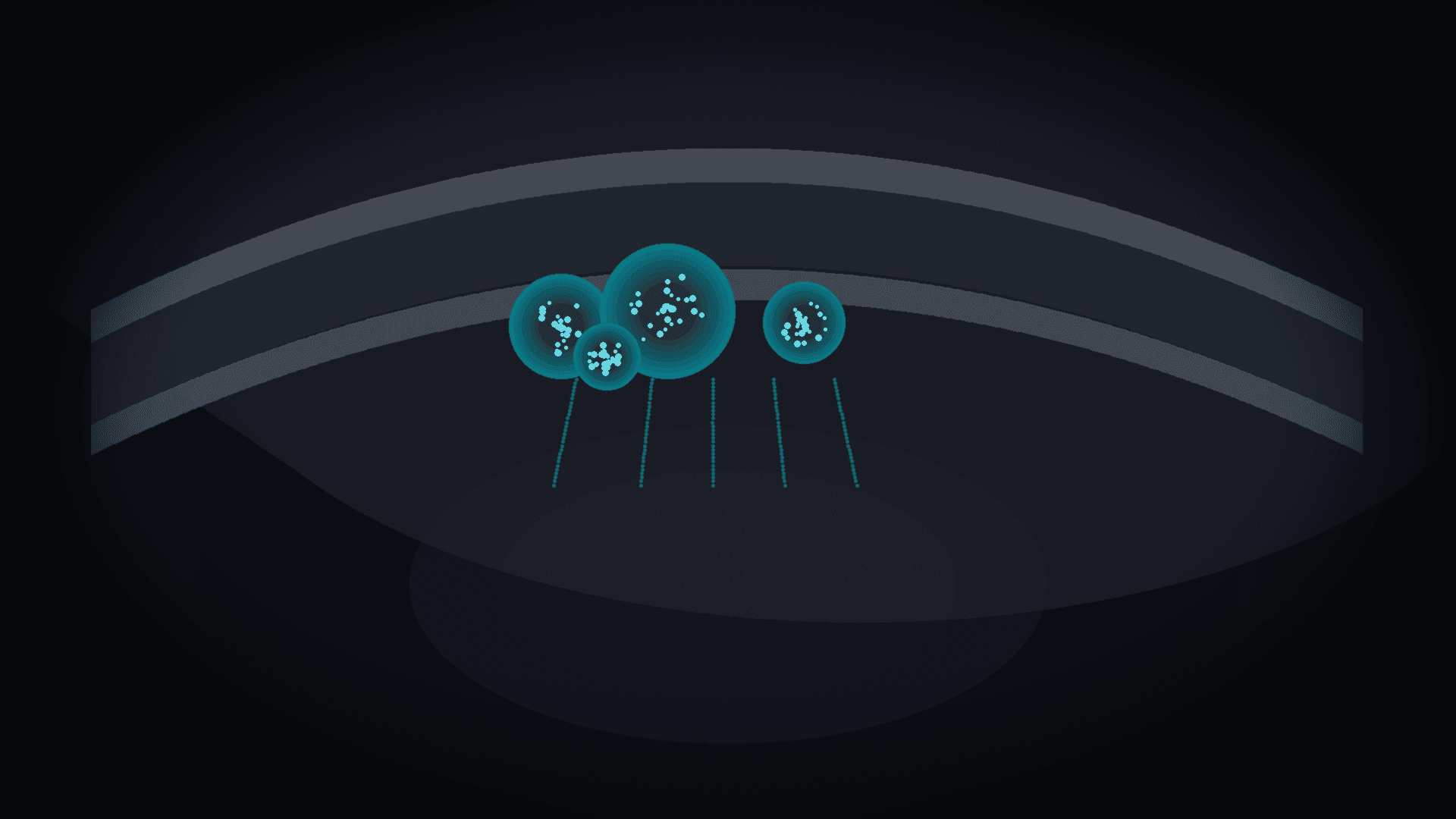
Picture a checkpoint at a border. Not on a highway and not in a capital. Right next to the building it protects. A neck lymph node is far from the brain. A team at Washington University School of Medicine in St. Louis shows in Nature that mice have a closer control point: lymphoid structures in skull bone marrow. This is not a slide-deck metaphor. It is anatomy and function, measured in the lab.
The paper appeared on 19 August 2026 (DOI: 10.1038/s41586-026-10951-4, CC BY 4.0). First author: Jang Hyun Park; corresponding authors: Park and Jonathan Kipnis. The authors identify skull-marrow clusters that resemble lymph-node germinal centres, with follicular-helper-like T cells (TFH). Those cells help B cells through CD40L, IL-21 and interferon-gamma signalling. The structures sense CNS-derived antigens and take part in anti-tumour responses in mouse brain-cancer models.
Scene: channels instead of a wall
For decades, textbooks taught that the brain is “immune privileged”: separated from blood by a barrier, without classical lymphatics, as if cut off from the body. Kipnis and others already showed that the meninges have lymphatic vessels, and that bony channels run between skull and dura. Through those channels, cerebrospinal fluid and immune cells exchange with skull marrow — in health and disease.
The new scene is the next step. Skull marrow is not only a blood-cell warehouse. The authors find local “training stations” of adaptive immunity. Neck nodes still matter. The brain also has its own, closer outpost.
In the WashU Medicine release, Park says such structures had not been seen before in healthy bone marrow. Kipnis adds that skull marrow is more than scaffolding — it harbours hubs for brain-specific immune responses.
Anatomy of the outpost
The authors compare skull marrow with sternum and femur marrow. TFH cells (including IL-21+CXCR5+) are clearly enriched in the skull. T–B clusters sit especially in posterior calvarial regions: occipital and interparietal. That matters: not “anywhere in the skull,” but closer to hindbrain routes and CSF outflow.
Those clusters show germinal-centre features: B-cell activation, antibody-secreting cells, and dependence on the CD40–CD40L axis and IL-21. When the team placed a hydrogel with a CD40L-blocking antibody under the scalp, the skull-marrow response weakened. The same gel on the flank did not produce that local effect. In mice, at least, the skull outpost can be targeted without shutting down the whole peripheral immune system at once.
Brain antigens reach marrow through channels and glymphatic-dependent flow. Aqp4-deficient mice, with impaired perfusion, took up less antigen in dura and skull marrow, mounted weaker local responses, and fared worse in the CT2A tumour model. Damaging deep cervical lymph nodes did not fully switch off this local route. The scene holds: the brain sends signals to nearby marrow, not only to distant nodes.
When a tumour appears in the brain
To test whether the outpost defends, the team used the CT2A glioma model. Local CD40L blockade in skull marrow via scalp hydrogel shortened survival. Lymphocyte infiltration and CD8 activation in the tumour also fell. That is not proof that “a skull node cures cancer.” It is proof that the niche matters in this model.
Then they pushed the other way: enhancement. A mix of CD40 agonist with IL-21 and interferon-gamma in a scalp hydrogel boosted local lymphoid responses in skull marrow and prolonged survival. Local CD40 agonist alone could be harmful (earlier mortality, possibly regulatory B cells). The three components together worked better. Responses rose first in skull marrow, later in nodes outside the skull — “locals first, reinforcements later.”
Local treatment still worked after craniotomy-free tumour injection and in mice with compromised classical secondary lymphoid organs. Efficacy depended on B cells and antibody responses. That bounds the marketing fantasy: not a miracle glioma gel, but local boost of a defined niche, in mice, in one model.
A human clue, not a drug
Human skull-marrow data (dataset GSE233304) support TFH-like cells. That is a bridge, not a highway to the clinic. There is no Phase I trial, no patient dose, no approval. In the release, Kipnis lists immune-component diseases — from Alzheimer’s to long COVID — as a horizon for skull-directed therapy with fewer peripheral side effects. That is a research hypothesis, not a product promise.
We put this story at TRL about 2–3. The anatomical–functional discovery level is high (Nature, multi-modal evidence). The therapeutic technology level is low: mouse models, subcutaneous gel, no clinical validation. Barriers are clear: mouse–human anatomy differences, CD40-agonist safety, autoimmunity risk when “turning up” immunity near the brain, scalp delivery in people, and regulation.
What we already knew — and what was missing
Skull channels and CSF contact with marrow were described earlier. Skull marrow was known to supply immune cells to CNS borders. Missing was whether adaptive responses specific to CNS antigens occur there, with their own training structures.
Park and Kipnis answer yes. Skull marrow shows not only scattered lymphocytes but organisation: clusters, T–B interaction, dependence on helper axes (CD40L, IL-21, interferon-gamma). A cell warehouse is one thing. A local immune school is another.
They also checked that results were not artefacts of skull surgery. Tumour injection via the cisterna magna without craniotomy reproduced key findings.
Scene at cell scale
Immunofluorescence shows B220 (B), CD4 (T) and IL-21 clusters in posterior vault regions. Single-cell and flow analyses mark TFH cells with Pdcd1, Cxcr5, Bcl6, Il21 and proteins CD40L, ICOS and BCL6. Sternum and femur carry less of this signature. Posterior skull stands out in TFH counts and cluster area.
When CT2A appears, germinal-centre-like B cells rise in skull marrow. The local response comes earlier than full expansion in distant nodes — first responders beside, not instead of, cervical nodes.
Limits of the evidence
The paper does not claim humans host a ready-made lymphatic organ identical to the mouse map. It shows TFH-like cells in human skull marrow and coherent mouse functional data. We do not invent survival percentages from unread plot ticks.
Nor is the scalp hydrogel a finished drug platform. In the study it is a tool: local delivery of antibodies and cytokines in carboxymethylcellulose hydrogel. Human pharmacokinetics, scalp infection risk and dose repeatability remain open.
Business layer, separate
Science says: a local niche exists. Business and clinic ask: who pays for a skull-marrow-directed therapy, and when. Today: nobody buys a finished product from this paper. Kipnis co-founded Pranas Neuro, Rho Bio and Argo Neurotech — a signal that neuroimmune spin-offs are forming, not that the Nature gel launches in 2027.
Investors and hospitals care about path: biomarker (is the niche active in a patient?), route (scalp vs invasive), safety (CD40, cytokines), indication (gliomas? neurodegeneration? MS?). Each is a separate programme. Jumping from Nature to “Alzheimer’s drug” is a communication error we avoid here.
Poland and Europe angle
Europe has strong neuro-oncology and neuroimmunology centres; Poland has glioma clinics, MS centres and growing meningeal and blood–brain-barrier labs. A sensible multi-year scenario is not “Polish CD40 gel,” but joining networks that map human skull channels, test safer local immunomodulators and build imaging or molecular readouts of niche activity.
For EU health policy the theme is double. First: why systemic therapies poorly reach the brain or hit the whole body. Second: skull-local targets might cut peripheral exposure — if they ever clear validation. That is a research argument, not a budget miracle.
What the scene changes for the reader
The main shift is not “we have a drug.” It is: the brain is not a lonely island. It has a nearby outpost in the bone that wraps it. When antigen leaves the brain, local lymphoid structures can react before a neck node. When that outpost is blocked, mice in a glioma model fare worse. When it is locally boosted with three signals, they live longer — under experimental conditions.
The scene can be told without grant jargon. Bone. Marrow. Cell cluster. Channel to dura. Scalp gel as a lab tool, not a pharmacy cream. A human clue in expression data. And an open question: will this anatomy someday change how neurological therapies are designed?
For today, Nature documents the fact: mouse skull bone marrow hosts lymphoid structures that survey the CNS. The outpost is closer to the brain than a classical neck node. Clinic, spin-offs and approvals are still being drawn.
Sources
- J. H. Park et al., Functional role of skull lymphoid structures in CNS immunosurveillance, Nature (2026). DOI: 10.1038/s41586-026-10951-4. CC BY 4.0.
- WashU Medicine, Newly found ‘immune organ’ inside skull directs brain defense, 19 August 2026. https://medicine.washu.edu/news/newly-found-immune-organ-inside-skull-directs-brain-defense/
Comments· 0
No comments yet. Be the first.