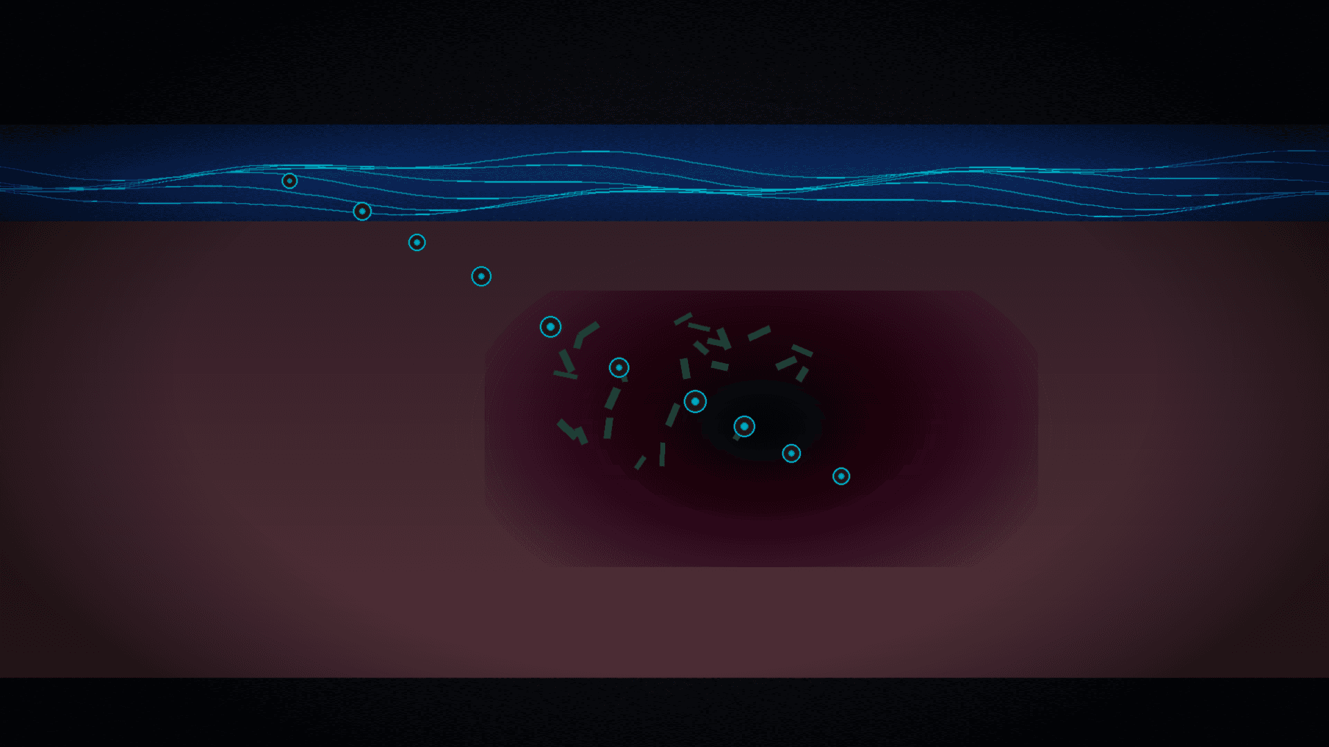
A nanocapsule delivers the antibiotic to the ulcer despite stomach mucus
The stomach looks like a simple acid-filled pouch. The anatomy of a Helicobacter pylori infection is more layered: acid, a mucus sheet, the floor of an ulcer, and only then bacteria hidden hundreds of micrometres deep. A standard clarithromycin tablet moves through that system like a letter dropped into the wrong mailbox: some of it dissolves too early, some sticks in mucus, and too little reaches the deep focus. The clinician then raises the dose, and resistance rises with it.
A team at Pusan National University led by Jin-Wook Yoo designed a layer-by-layer answer. In the Journal of Controlled Release (DOI: 10.1016/j.jconrel.2026.114869), Hyunwoo Kim and colleagues describe PLGA nanoparticles coated with polydopamine and loaded with clarithromycin. This is not another “smart carrier” slogan from a catalogue. It is a delivery cascade matched to the anatomy of an H. pylori–infected gastric ulcer.
Why half of humanity still loses to one bacterium
H. pylori affects about half of the world’s population. It is linked to peptic ulcers and higher gastric-cancer risk, and after decades of eradication regimens it remains everyday clinic work. Antibiotics are the standard, but “standard” does not mean the drug physically reaches where the pathogen sits.
The bacterium uses organ topography: it shelters under mucus and inside altered ulcer tissue. Systemic therapy then has to punch through barriers that evolution built against acid, not against pharmacists. The result is predictable — higher doses, more adverse effects, stronger selection for resistant strains. The Pusan authors stated the need plainly: systems that deliver the antibiotic locally, instead of flooding the whole body in the hope that a fraction of the dose hits the target.
Layer 1: acid that destroys the drug
The first anatomical layer is chemistry, not microbiology. Low gastric pH breaks many molecules before they can act. The platform’s core was made from biodegradable PLGA by nanoprecipitation, then coated with polydopamine — a material known for surface adhesion chemistry, inspired in part by mussel proteins.
Polydopamine is not a marketing ornament here. In physicochemical characterisation (size, morphology, surface chemistry, stability, drug loading and release profile), the platform stayed stable under acidic conditions and limited premature clarithromycin release. The first layer stops being the place where the drug dies before it starts working. That sentence is banal and, at the same time, the boundary condition of the whole cascade: without surviving acid, talking about mucus or ulcer depth is pointless.
Layer 2: mucus as a curtain
H. pylori does not sit on the surface like a bacterium on a kitchen counter. It hides under mucus — a gel barrier that protects the stomach wall from its own acid and, as a side effect, blocks antibiotic access. Raising the systemic dose is an attempt to smash the curtain with force rather than a key.
In in vitro and ex vivo studies the nanoparticles penetrated the mucus barrier. That step does not appear on a package leaflet, yet it decides whether the molecule ever meets the bacterium. Without mucus penetration the rest of therapy is a pharmacokinetic lottery: the drug circulates, the patient waits, and the pathogen stays on its own floor of the building.
This is the difference between “nanomedicine as a slogan” and nanomedicine as transport engineering. Particle size alone is not enough. What matters is whether surface and hydrodynamics let the particle cross the mucus gel instead of sticking in it like flies on tape.
Layer 3: the ulcer as an address, not scenery
An ulcer is not only a “hole in the mucosa”. It is a local inflammatory focus, altered tissue, and a place where H. pylori may sit deeper than an endoscopic image suggests. The Pusan platform was designed to accumulate selectively at ulcer sites and prolong gastric retention — to stay there longer instead of washing downstream with gastric contents.
Anatomy becomes logistics: “drug in the stomach” is not enough. You need drug in a specific millimetre of altered wall, for long enough for the antibiotic to act locally. Retention is an underrated parameter in eradication debates. You can have an excellent antibiotic and a fatal contact time with the target.
Layer 4: four hundred micrometres deep
The strongest detail from the paper and the Pusan/phys.org account: the nanoparticles reached about 400 μm into ulcer tissue — where deep-seated bacteria reside. That scale is invisible to the naked eye at gastroscopy, and it helps explain why “general” therapies often fail in patients with ulcers.
Polydopamine also enabled strong, ligand-independent adhesion to bacteria. Instead of hunting for a single molecular key, the coating provided contact and local clarithromycin release directly at the pathogen. In a mouse model of H. pylori–infected gastric ulcers the authors report roughly 99.9% bacterial reduction, faster ulcer healing, longer retention and support for tissue regeneration — at a clarithromycin dose tenfold lower than conventional systemic therapy in that comparison.
Ten times less antibiotic with preserved or better local efficacy is a clinical, economic and epidemiological argument at once. Lower whole-body exposure means fewer side effects. A lower systemic dose means weaker selection for resistance. For the patient, a simpler regimen can be the difference between finishing therapy and abandoning it halfway.
What the lab actually did
The work does not stop at a coating idea. The authors characterised the nanoparticles for size and morphology, surface chemistry, stability, drug loading and release, then ran a validation chain: in vitro tests, ex vivo preparations and a mouse model of H. pylori–infected gastric ulcers. Along that chain they assessed mucus penetration, bacterial adhesion, gastric retention, tissue penetration, antibacterial activity and therapeutic efficacy.
That study layout matters for TRL scoring. “Nanoparticle + antibiotic” in a tube is usually TRL 2. Here the platform crosses biological barriers in an animal model with measurable effect (bacterial reduction, healing, lower dose). That pushes the story into the TRL 3–4 neighbourhood — still before humans, but already beyond a pure materials idea.
It also helps separate facts from popular narrative. The figures 400 μm, 99.9% and 10× come from the Pusan/phys.org account of the publication; the full Elsevier text is closed (no CC BY licence for figures), so this TRL09 article does not copy publisher illustrations. The hero is a conceptual image of the anatomical cascade, not a reproduction of a paper figure.
From mouse to shelf: an honest TRL
Technology readiness: about TRL 3–4. There are in vitro, ex vivo and in vivo mouse data; there are no human clinical trials, no pharma-grade manufacturing scale-up, and no proof that the same profile holds in a human stomach with meals, acid-suppressing drugs and comorbidities.
Commercial barriers are predictable and worth listing without wrapping:
- manufacturing nanoparticles to pharmaceutical quality (batch repeatability, sterility, impurity control);
- stability in acid and under post-meal pH swings;
- head-to-head eradication rates versus standard of care in clinical studies;
- intellectual-property position versus other mucoadhesive carriers and polydopamine coatings;
- clinical development spend — costlier than another combination of known tablets, potentially cheaper per patient in antibiotic use if the local dose holds.
In the university account, Jin-Wook Yoo stresses that a targeted strategy could simplify treatment by cutting the required dose while keeping efficacy — with possible gains in adherence and fewer treatment failures. He also notes that the local-delivery platform could later be adapted to other diseases where biological barriers blunt conventional drugs. That is a technology-platform preview, not a promise of a finished product on the shelf.
A Poland angle without illusions
In Poland H. pylori is not an exotic university-clinic topic — it is everyday gastroenterology and primary care. Eradication regimens generate costs, adverse effects and relapses when local resistance is high. A nanoparticle courier from Korea will not replace society guidelines tomorrow. It can, however, point the direction when assessing spin-offs and drug-delivery projects: not another antibiotic analogue, but an anatomically designed carrier that cuts the systemic dose.
For a deep-tech investor the question is not whether PLGA and polydopamine are new — they are not. It is whether the full cascade (acid stability → mucus penetration → ulcer retention → bacterial adhesion → local release) yields a measurable eradication edge at a lower dose, and whether that can be reproduced in manufacturing. Today the answer is: in the mouse model — yes; in the clinic — not yet known.
Polish labs and pharma firms know formulations and generics. The gap is not only in synthesising another molecule, but in engineering delivery to where the pathogen actually lives. The Pusan work is therefore a market signal: value may form at the junction of polymer materials and gastroenterology, not only in a new antibiotic mechanism of action.
In practice, investor and clinician get the same checklist: will the nanoparticle survive acid, cross mucus, stay in the ulcer, reach hundreds of micrometres deep, and do so at a meaningfully lower dose than today? Until answers pass through human studies, the story remains a promise with a map — better than a promise without one, but still a promise.
Anatomy instead of a slogan
This story’s mode is not a duel between two firms or a materials paradox. It is the anatomy of a drug’s path. Each stomach layer filters out a different weakness of classical therapy. The Pusan nanoparticle does not kill bacteria by “nanotech magic”; it passes the filters that used to lose clarithromycin.
If later stages — toxicology, pharmacokinetics in larger animals, first human studies — confirm the mouse profile, gastroenterology may get a tool closer to precise local intervention than to another catch-all tablet. For now we have a carefully built cascade and numbers worth remembering: 400 μm into tissue, 99.9% bacterial reduction in the model, a 10× lower dose. The rest is the road from TRL 3–4 to the pharmacy shelf — long, but this time with an anatomical map instead of guesswork.
Sources
- Hyunwoo Kim et al., Polydopamine-mediated multi-stage delivery for precise local therapy of Helicobacter pylori–infected gastric ulcers, Journal of Controlled Release (2026). DOI: 10.1016/j.jconrel.2026.114869
- Pusan National University / phys.org, Smart nanoparticles deliver antibiotics directly to H. pylori (2026-09-15): https://phys.org/news/2026-09-smart-nanoparticles-antibiotics-pylori.html
- Crossref record for DOI 10.1016/j.jconrel.2026.114869 (authors; funder National Research Foundation of Korea / Ministry of Science and ICT; award NRF-2022R1A2C2004340)
Comments· 0
No comments yet. Be the first.