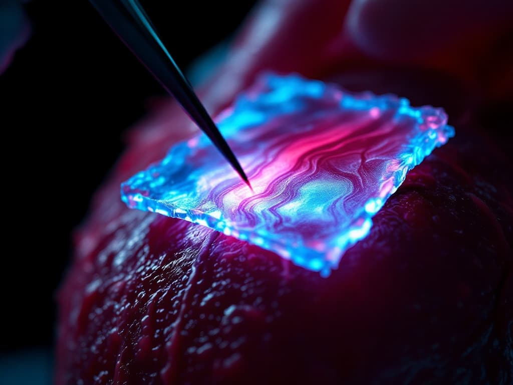
A billion cells vanish forever. A patch of living muscle changes that
A billion cells that disappeared
A heart attack lasts a few minutes, but its effects stay forever. When a clot blocks a coronary artery, a fragment of heart muscle — sometimes the size of a thumb — dies from oxygen starvation. As many as a billion cardiomyocytes are lost. These are the cells that contract eighty times a minute, without a single break, for eighty years.
And here the paradox begins. A cut liver regrows. Skin seals itself. Bone knits back together. The heart — the organ that works hardest and longest — cannot repair itself at all. A lost cardiomyocyte does not come back. In its place a scar grows that does not pump. It only stiffens.
For the patient this means heart failure: the muscle weakens, and for the most severe cases the only lasting rescue is a transplant. Except there are not enough donor hearts, and the waiting list grows faster than the list of donors.
For thirty years, science tried to work around this. And that is exactly why this story has a false hero.
First wall: the cell that refuses to divide
Why can the heart not repair itself at all? Because cardiomyocytes, unlike skin or liver cells, have almost completely abandoned the ability to divide. This is the price of precision: a heart muscle cell is so specialized for contracting that it lost the machinery needed for multiplying.
When in 1994 Mark Field's team at Indiana University implanted fetal cardiomyocytes into mouse hearts and saw under the microscope that donor cells linked up with recipient cells into a single synchronized muscle, the scientific world froze with hope. Science published it as a breakthrough. If you can implant foreign cells and make them beat together with the heart — then maybe you can replace the lost ones.
There was only one problem that nobody saw at the time.
False trail: inject and hope
Over the next two decades, the entire field bet on a single strategy. Simple as a needle. Grow millions of cardiomyocytes in the lab from stem cells — first embryonic, then induced pluripotent — and inject them straight into the wall of the damaged heart.
On paper, the results looked wonderful. In 2014, James Chong and Charles Murry from the University of Washington reported in Nature that human cardiomyocytes injected into macaques after a heart attack rebuilt a fragment of muscle. In 2016, Yuji Shiba's team showed the same with iPS cells. In 2018, another paper in Nature Biotechnology. Citations climbed. Companies formed. Clinical trials launched. Everything suggested the solution was within reach.
The data looked so good that few people asked the uncomfortable question: what exactly does an injected cell do once it lands in the heart wall?
Why it failed: they beat, but out of rhythm
The answer came in 2019, in a paper by Romagnuolo and colleagues whose title says it all: “Human embryonic stem cell-derived cardiomyocytes regenerate the infarcted pig heart but induce ventricular tachyarrhythmias”.
An injected muscle cell is like a musician who walks into an orchestra and starts playing his own melody. It beats — but at its own tempo. Millions of cells injected into the heart wall do not synchronize with the rest of the muscle. They generate chaotic electrical impulses. The result: ventricular rhythm disturbances that can kill a patient faster than the heart failure itself.
Then came the second problem — quantitative. Most injected cells died within the first few days. Survival at the level of a few percent meant the therapy was expensive and the effect meager. And the third problem, the most dangerous: if even one undifferentiated pluripotent cell remained among the injected cells, it threatened a teratoma — a tumor.
Three independent failures. One strategy that for twenty years looked like the only right one.
Breakthrough: a patch instead of an injection
Wolfram-Hubertus Zimmermann in Göttingen looked at the same problem from a different angle. His question was: why inject individual cells and hope they organize themselves — when you can first build an entire, beating sheet of muscle in the lab?
This is the difference between dumping a handful of bricks on a construction site and delivering a finished wall. Over the years, Zimmermann refined “engineered heart muscle” — EHM, patches of living heart muscle. As early as 2006 he showed in Nature Medicine that such a patch improves the function of a rat heart after a heart attack. In 2017 his team developed a version for humans, produced to GMP standards — the way a drug is manufactured.
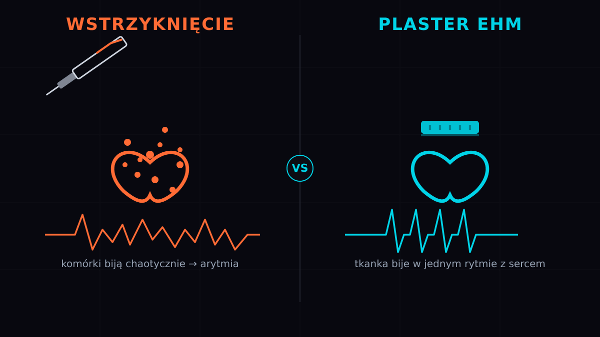
Rys. 1. Dwie strategie naprawy serca: wstrzyknięte komórki biją chaotycznie i wywołują arytmię, a łata EHM przyszyta do nasierdzia bije w jednym rytmie z sercem. Opracowanie własne TRL09.
The idea is simple to describe and devilishly hard to execute. From iPS cells you grow two cell types: cardiomyocytes and stromal cells, which give them a scaffold. You mix them in a ratio, culture them in special molds — and after a few weeks you get a piece of tissue that genuinely beats. You do not inject it into the heart wall. You sew it onto the surface — onto the epicardium, like a patch on a tire inner tube.
The key lies in maturation. Cells in a patch do not reach the heart raw — for weeks in a bioreactor they are stretched and mechanically stimulated, exactly like a working muscle. Before they reach the operating table, they learn to beat in a single rhythm. An injected cell has no such training.
In January 2025, Nature published the results. In macaques with chronic heart failure, EHM patches built from 40 to 200 million cells thickened the heart wall and improved contractility and ejection fraction. Crucially: in twenty implanted macaques — 66 patches, 2.4 billion cells in total — neither arrhythmia nor tumor was observed. The cells survived for at least six months.
Patient number 27016
But the paper's most important sentence concerns a human. Based on these data, the German regulator Paul-Ehrlich-Institut approved the world's first clinical trial of heart repair with engineered tissue — BioVAT-HF-DZHK20.
The first patient, designated number 27016, suffered from advanced heart failure. He received ten EHM patches — 400 million cells in total — and waited for a transplant. Three months later he received a new heart. And the old one, removed during the transplant, went under the microscope.
What the pathologists saw closed thirty years of debate. The EHM patches were visible on the surface of the heart with the naked eye. Under the microscope — donor cardiomyocytes that had survived, vascularized, and grown into the recipient's muscle. Remuscularization of the human heart, documented for the first time not in a model but in a human being.
This was not a single dose. The protocol called for escalation: from five patches in the macaque model — the maximum dose deemed safe — through ten patches in the first patient, up to twenty patches built from 800 million cells. Each step was a boundary no one had crossed before. And each one went the same way: no arrhythmia, no tumor.
The race nobody sees
The stakes are enormous. Heart failure affects over 60 million people worldwide, and the therapy market is counted in tens of billions of dollars. “Patch-on-heart” technology changes the rules of the game: it is not another attempt to replace cells, but a ready tissue product that can be standardized, stored, and implanted as part of routine cardiac surgery.
The competition is not asleep. The Japanese firm Heartseed is testing spheroids of iPS-derived cardiomyocytes. The American BlueRock Therapeutics, owned by Bayer, is working on injections of cardiomyocytes — with the same arrhythmia risk that buried the previous approach. The Göttingen team took a different path: the company Repairon GmbH commercializes EHM patches and runs the clinical trial.
The difference between the competitors boils down to a single sentence in Zimmermann's paper: injected cells fire impulses and cause arrhythmia, while an EHM patch does not. This is not a nuance. It is the difference between a therapy and a threat.
There is one more reason the patch beats the injection hands down — literally. Injected cells are a suspension: you cannot control where they land and how they arrange themselves. An EHM patch is a product with a defined shape, thickness, and contraction force. You can measure it, weigh it, and describe it in a drug dossier. And that is precisely the condition for a regulator to approve the therapy at all, and for a hospital to order it like any other medical device.
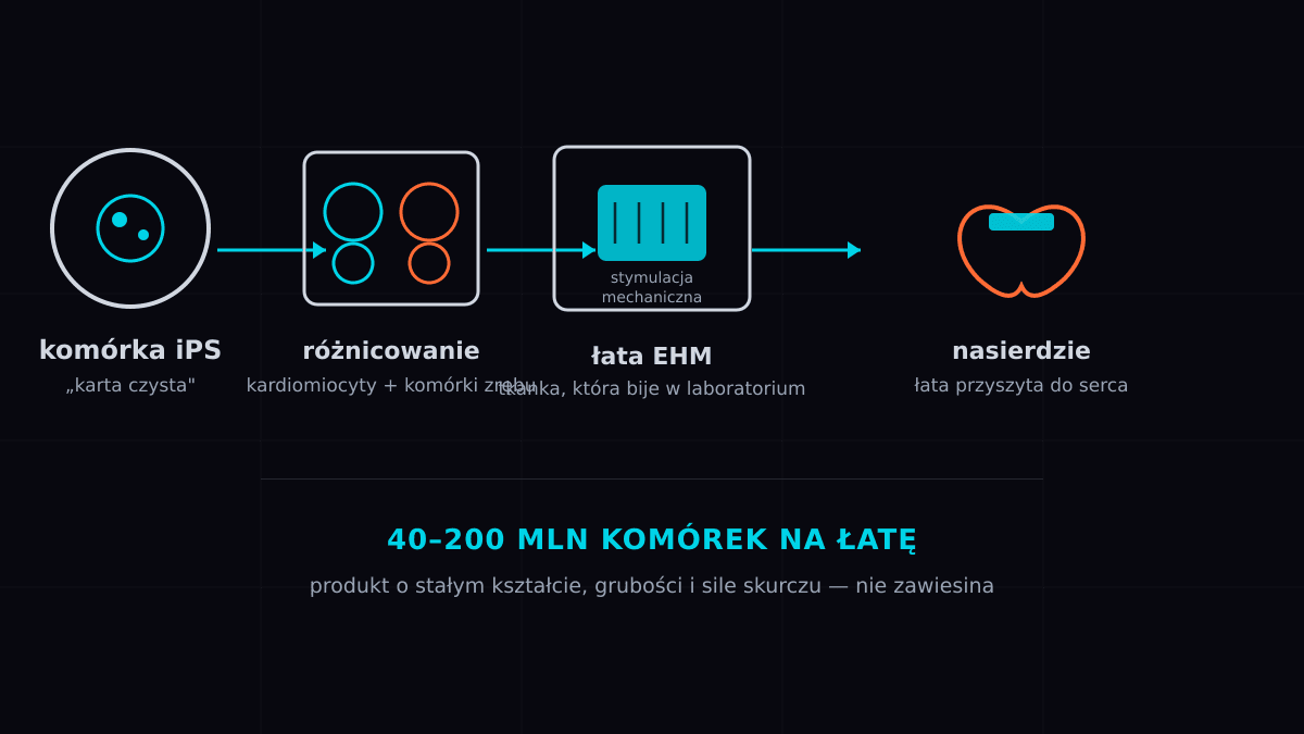
Rys. 2. Produkcja łaty EHM: komórki iPS różnicują się w kardiomiocyty i komórki zrębu, dojrzewają pod stymulacją mechaniczną, a gotową łatę przyszywa się do nasierdzia. Opracowanie własne TRL09.
Poland: a transplant heart waiting for a patch
For Poland this technology carries exceptional weight. Cardiovascular disease is our leading cause of death, and heart failure affects about a million Poles. At the same time, the Silesian Center for Heart Diseases in Zabrze ranks among Europe's leading heart transplant centers — Polish cardiac surgeons perform around two hundred transplants a year, but the waiting list is always longer than the list of donors.
And this is exactly where the “patch” comes in. BioVAT-HF is testing EHM as a bridging therapy — a bridge to transplant. A patient who today dies waiting for a donor could receive a patch from their own cells and live to reach the transplant in better shape. The scale of the problem is concrete: in Poland the wait for a heart transplant often exceeds a year, and some patients do not make it. A therapy that buys a patient a few extra months without arrhythmia risk would change survival statistics from day one. For a country with such advanced transplantology and such long queues, this is not a curiosity but a real gap to fill.
Poland also has the infrastructure not to stand on the sidelines of this race. FamiCord (formerly the Polish Stem Cell Bank) is one of the largest stem cell banks in Europe — the infrastructure for storing and expanding iPS cells already exists. The Medical Research Agency funds non-commercial clinical trials, and Polish cardiac surgery centers have experience that many Western clinics lack. What is missing is one thing: a Polish center that would take on producing EHM patches and join a clinical trial.
The story has come full circle. Thirty years ago everyone believed in the syringe. Today we know the heart is repaired not with an injection but with a patch — patiently, cell by cell, in the laboratory, before it reaches the operating table. The first human already wore one. The next ones are waiting.
Sources
- Jebran A.-F. i in., Engineered heart muscle allografts for heart repair in primates and humans, Nature (2025). DOI: 10.1038/s41586-024-08463-0
- Romagnuolo R. i in., Human embryonic stem cell-derived cardiomyocytes regenerate the infarcted pig heart but induce ventricular tachyarrhythmias, Stem Cell Reports (2019). DOI: 10.1016/j.stemcr.2019.04.005
- Chong J.J.H. i in., Human embryonic-stem-cell-derived cardiomyocytes regenerate non-human primate hearts, Nature (2014). DOI: 10.1038/nature13233
- Shiba Y. i in., Allogeneic transplantation of iPS cell-derived cardiomyocytes regenerates primate hearts, Nature (2016). DOI: 10.1038/nature19815
- Zimmermann W.-H. i in., Engineered heart tissue grafts improve systolic and diastolic function in infarcted rat hearts, Nature Medicine (2006). DOI: 10.1038/nm1394
- Tiburcy M. i in., Defined engineered human myocardium with advanced maturation for applications in heart failure modeling and repair, Circulation (2017). DOI: 10.1161/CIRCULATIONAHA.116.024145
- Soonpaa M.H. i in., Formation of nascent intercalated disks between grafted fetal cardiomyocytes and host myocardium, Science (1994). DOI: 10.1126/science.8140423
Comments· 0
No comments yet. Be the first.